There are two types of cell division: mitosis and meiosis. Most of the time when people refer to “cell division,” they mean mitosis, the process of making new body cells. Meiosis is the type of cell division that creates egg and sperm cells.
Mitosis
Mitosis is the type of cell division by which a single cell divides in such a way as to produce two genetically identical "daughter cells". This is the method by which the body produces new cells for both growth and repair of aging or damaged tissues throughout the body. Mitosis is also referred to as "binary fission".
Meiosis
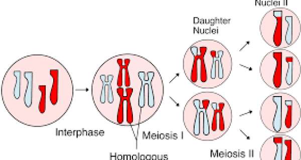
Meiosis, which is also referred to as "reduction division", is the form of cell division in which a cell divides into four "daughter cells" each of which has half** of the number of chromosomes of the original cell. Meiosis occurs prior to the formation of sperm (in males) and ova (in females). That is - meiosis only occurs in the "gametes".
**The cells return to having the normal (called "diploid") number of chromosomes after fertilization of the ova by the sperm.
Meiosis consists of two successive divisions, each of which is divided into four phases. The first meiotic division is similar to mitosis (defined above) and the second meiotic division is the "reduction" stage. Meiosis enables the exchange of genetic material between chromosomes.
Mitosis Is Divided into Well-Defined Phases
mitosis is understood to involve five phases, based on the physical state of the chromosomes and spindle. These phases are prophase, prometaphase, metaphase, anaphase, and telophase. Cytokinesis is the final physical cell division that follows telophase and is therefore sometimes considered the sixth phase of mitosis.
Prophase
Mitosis begins with prophase, during which chromosomes recruit condensin and begin to undergo a condensation process that will continue until metaphase. In most species, cohesin is largely removed from the arms of the sister chromatids during prophase, allowing the individual sister chromatids to be resolved. Cohesin is retained, however, at the most constricted part of the chromosome, the centromere. During prophase, the spindle also begins to form as the two pairs of centrioles move to opposite poles and microtubules begin to polymerize from the duplicated centrosomes.
Prometaphase
Prometaphase begins with the abrupt fragmentation of the nuclear envelope into many small vesicles that will eventually be divided between the future daughter cells. The breakdown of the nuclear membrane is an essential step for spindle assembly. Because the centrosomes are located outside the nucleus in animal cells, the microtubules of the developing spindle do not have access to the chromosomes until the nuclear membrane breaks apart.
Prometaphase is an extremely dynamic part of the cell cycle. Microtubules rapidly assemble and disassemble as they grow out of the centrosomes, seeking out attachment sites at chromosome kinetochores, which are complex platelike structures that assemble during prometaphase on one face of each sister chromatid at its centromere. As prometaphase ensues, chromosomes are pulled and tugged in opposite directions by microtubules growing out from both poles of the spindle, until the pole-directed forces are finally balanced. Sister chromatids do not break apart during this tug-of-war because they are firmly attached to each other by the cohesin remaining at their centromeres. At the end of prometaphase, chromosomes have a bi-orientation, meaning that the kinetochores on sister chromatids are connected by microtubules to opposite poles of the spindle.
Metaphase
Next, chromosomes assume their most compacted state during metaphase, when the centromeres of all the cell's chromosomes line up at the equator of the spindle. Metaphase is particularly useful in cytogenetics because chromosomes can be most easily visualized at this stage. Furthermore, cells can be experimentally arrested at metaphase with mitotic poisons such as colchicine. Video microscopy shows that chromosomes temporarily stop moving during metaphase. A complex checkpoint mechanism determines whether the spindle is properly assembled, and for the most part, only cells with correctly assembled spindles enter anaphase.
Anaphase
The progression of cells from metaphase into anaphase is marked by the abrupt separation of sister chromatids. A major reason for chromatid separation is the precipitous degradation of the cohesin molecules joining the sister chromatids by the protease separase.
Two separate classes of movements occur during anaphase. During the first part of anaphase, the kinetochore microtubules shorten, and the chromosomes move toward the spindle poles. During the second part of anaphase, the spindle poles separate as the non-kinetochore microtubules move past each other. These latter movements are currently thought to be catalyzed by motor proteins that connect microtubules with opposite polarity and then "walk" toward the end of the microtubules.
Telophase and Cytokinesis
Mitosis ends with telophase or the stage at which the chromosomes reach the poles. The nuclear membrane then reforms, and the chromosomes begin to decondense into their interphase conformations. Telophase is followed by cytokinesis or the division of the cytoplasm into two daughter cells. The daughter cells that result from this process have identical genetic compositions.